Common Pigmentary Disorders - Focus on the Indian Scenario
Pigmentary disorders cause psychological distress and negatively impact the quality of life of an individual. Three important hyperpigmentary disorders in India, namely, melasma, postinflammatory hyperpigmentation (PIH) and actinic lentigines is detailed below.
Melasma Melasma, an acquired pigmentary disorder is characterized by hyperpigmented brown to grayish brown macules on the face. It occurs mainly in women (90% cases) and 10% of males of all ethnic and racial groups. In India, 20—30% of 40-65 years old women present a facial melasma. The current classification of melasma is based on the site of lesion and on the depth of pigmentation that is determined histologically or instrumentally within the epidermis, dermis or both.
While the exact cause of melasma is currently unknown, exposure to UV, increased estrogen levels (observed mainly during pregnancy or use of oral contraceptives), genetic predisposition and phototoxic drugs are known to play a major role in the development of this hypermelanosis disorder. Other factors implicated in its etiology include ovarian dysfunction, thyroid and/or liver diseases. The role of UV exposure has been shown to being crucial in development but mainly exacerbation of melasma. Epidemiological studies revealed that in more than 25% of cases, an association with sun exposure has been declared.
At the molecular level, it is well- established that exposure to UV rays induce increased production of alpha- melanocyte-stimulating hormone and corticotrophin as well as interleukin (IL)-1 that, in turn, contribute to increased melanin production. In addition, overexpression of dermal stem cell factor and its receptor, c- kit, have been identified in melasma lesions and are believed to increase melanogenesis. More generally in melasma, paracrine melanogenic factors have been identified from keratinocytes, mast cells or dermal fibroblasts. However, it seems that melasma results from complex interactions of various causative factors, UV exposure(s) included.

Actinic Lentigines
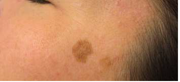
Actinic lentigines also called solar lentigines or lentigo senilis, are light brown to dark brown, spots, even-colored or reticulated patches occurring mainly in sun-exposed areas. The dorsal aspects of the hands, extensor forearms, upper trunk and face are the most commonly affected sites. They might be sometimes solitary, but these lesions are more often multiple. Actinic lentigines are considered to be a clinical sign of photoaging and their frequency increases with age. Their number is an indicator of the amount of sun exposure over the course of a life-time and therefore shows an increased risk for developing skin cancers.
Why do it in the winter IPL can be done any time of year, but it is ideal in the winter because most people spend less time outdoors. Sunlight can lead to possible darkening of treated spots if sun avoidance isn’t practiced and rosacea is usually worsened by sun exposure.
What it does This treatment improves the cosmetic appearance of sun damaged skin while also killing any pre-cancerous cells. A medication is applied to the entire face and soaks in for around an hour. “This allows any sun damaged skin and pre-cancerous cells to ‘suck up’ the medicine, while the normal cells do not. Then shine LED lights at the face which activates the medicine, killing the damaged cells that absorbed the photosensitizer medication. This seeks out not only visibly damaged cells, but also microscopically small pre-cancers that haven’t even shown up on the skin yet.
. They might be sometimes solitary, but these lesions are more often multiple. Actinic lentigines are considered to be a clinical sign of photoaging and their frequency increases with age. Their number is an indicator of the amount of sun exposure over the course of a life-time and therefore shows an increased risk for developing skin cancers. They are more characteristic of fair to the medium photoaged skin, and prevalence in India is quite close to other Asian countries. It affects one-third of women about 5O years old in the large sample study carried out in India and concerns half of the population over 7O years old. Actinic lentigines are sometimes difficult to distinguish from seborrheic keratosis or simplex lentigos. The presence of the three entities often causes distress to the individual due to their color, number, size, location, and their known link to aging for two of them.
. At the histological level, actinic lentigines are characterized by a hyperpigmented basal layer which is due to an increased total melanin content of the epidermis (hypermelaninosis). The number of melanocytes was found increased in some studies (hypermelanocytosis), whereas others did not reveal any differences. The global architecture of the epidermis is often disorganized with broadened rete-ridges and includes long, short, crowded, and bulbous buds. In addition, the rete-ridges of the dermal-epidermal junction are altered and elongated resulting in a protrusion of the epidermis into the dermis. Although a strong association between actinic lentigines development and UV exposure has been observed, the underlying molecular mechanisms are still not fully understood. a potential role of keratinocyte growth factor has been recently suggested as well as the involvement of other dermal factors.
